
Clinical history
In December 2025 we received the case of Rita, a donkey with chronic laminitis of many years' duration. As a rescued animal, there was no clinical history or previous radiographic study that could reveal the origin or progression of the disease.
Initial assessment
A complete clinical examination was performed together with a radiographic study to determine the severity of the pathology.
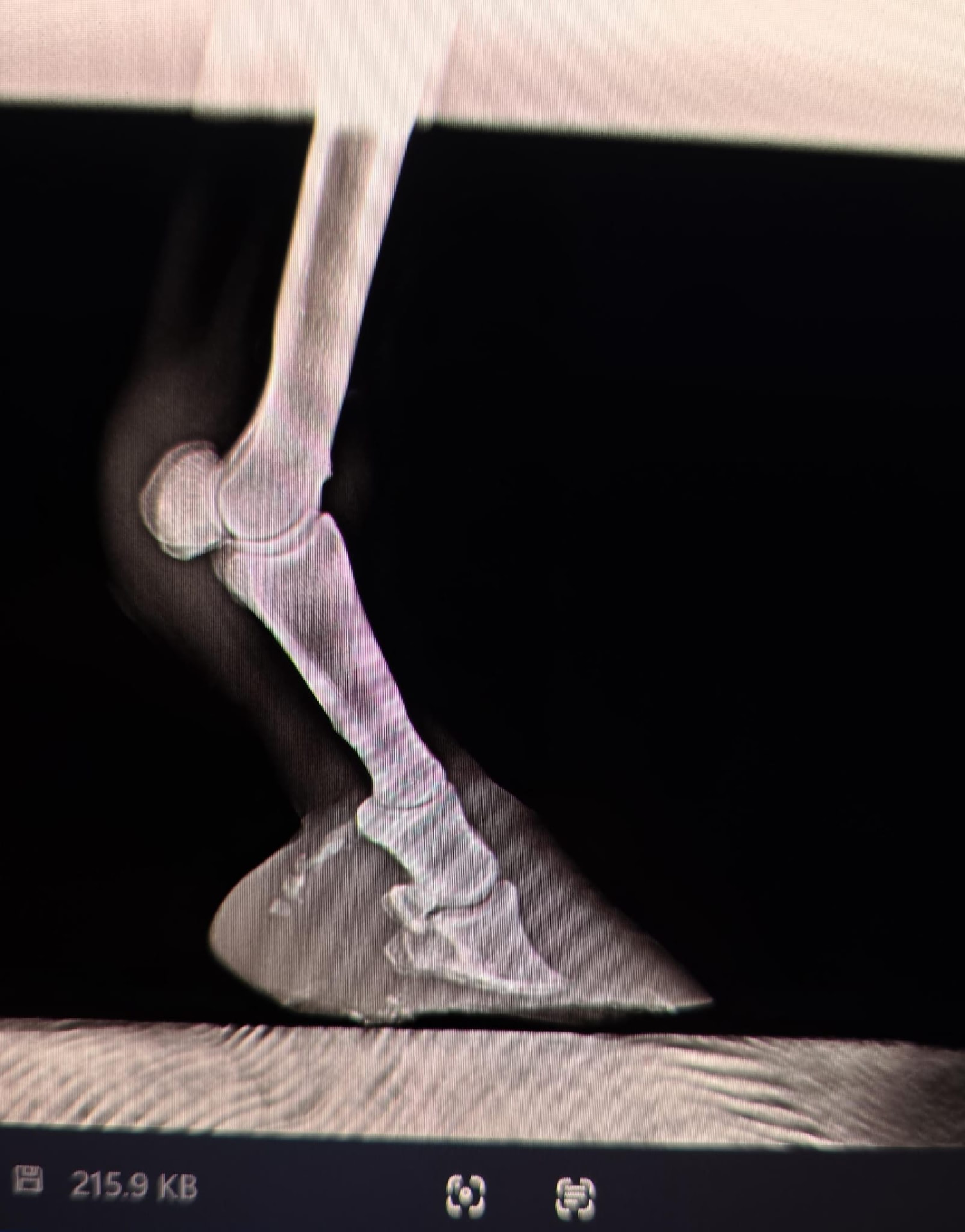
The initial radiographs revealed very severe alterations of the distal phalanx (P3), including marked bone remodelling, significant loss of bone mass and extensive osteolysis.
The remodelling of P3 was the consequence of years with a sole thickness insufficient to provide adequate support to the distal phalanx. As a result, P3 exerted excessive, constant pressure on the solar corium, the highly vascularised tissue responsible for sole production.
This sustained compression compromised the blood supply to the solar corium, preventing its normal function and limiting the production of new sole. Consequently, the distal phalanx remained very close to the ground, establishing a vicious circle in which the lack of sole depth allowed compression of the solar corium to continue, further reducing its capacity to produce horn tissue.
The characteristic curvature observed in P3 was the result of years of mechanical overload and bone remodelling. Likewise, the significant loss of bone mass seen radiographically was the consequence of a chronic reduction in blood supply to the bone.
The initial radiographs also showed a sole prolapse, where the distal phalanx displaced the solar tissue outwards due to the absence of sufficient sole depth to absorb load.
Treatment
3D-printed horseshoes were designed and manufactured specifically for this case with the aim of restoring blood supply to the solar corium.
The therapeutic principle was to reduce the tension exerted by the deep digital flexor tendon (DDFT) on the distal phalanx (P3), thereby decreasing the pressure applied to the solar corium. By reducing this compression, blood flow to the corium was restored, allowing its physiological function to recover and sole production to resume.
Treatment included periodic corrective trimming and replacement of the horseshoes as the case progressed, adapting hoof biomechanics to the needs of each stage of recovery.
Results
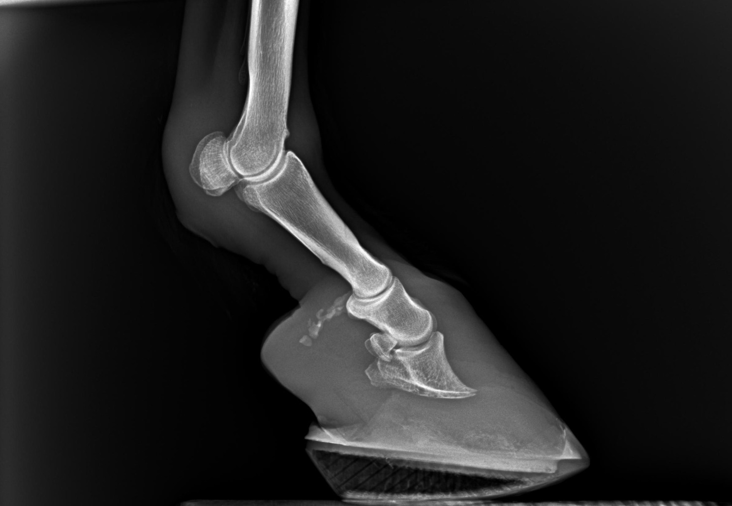
As a result of the treatment, sole thickness was at least tripled. This increase in solar support allowed a progressive improvement in the position of the distal phalanx (P3), reducing tension on the dermal laminae and favouring their repair.
Progression was confirmed radiographically through increased sole depth and the progressive repositioning of P3. In parallel, during the periodic trims a gradual recovery of the white line was observed, moving from a wide, pathological separation towards its normal morphology, presenting as a thin, continuous line with small yellowish areas consistent with tissue undergoing reorganisation and healing.
Clinically, Rita experienced a very significant improvement in comfort, showing greater willingness to move and an evident improvement in her quality of life.