
Clinical history
Mokka, an adult pony with an approximately two-year history of chronic laminitis, was referred to our service for assessment and treatment.
Clinically, Mokka presented a grade 3/5 lameness at the walk together with marked difficulty turning, showing evident functional limitation and signs of pain associated with the pathology.
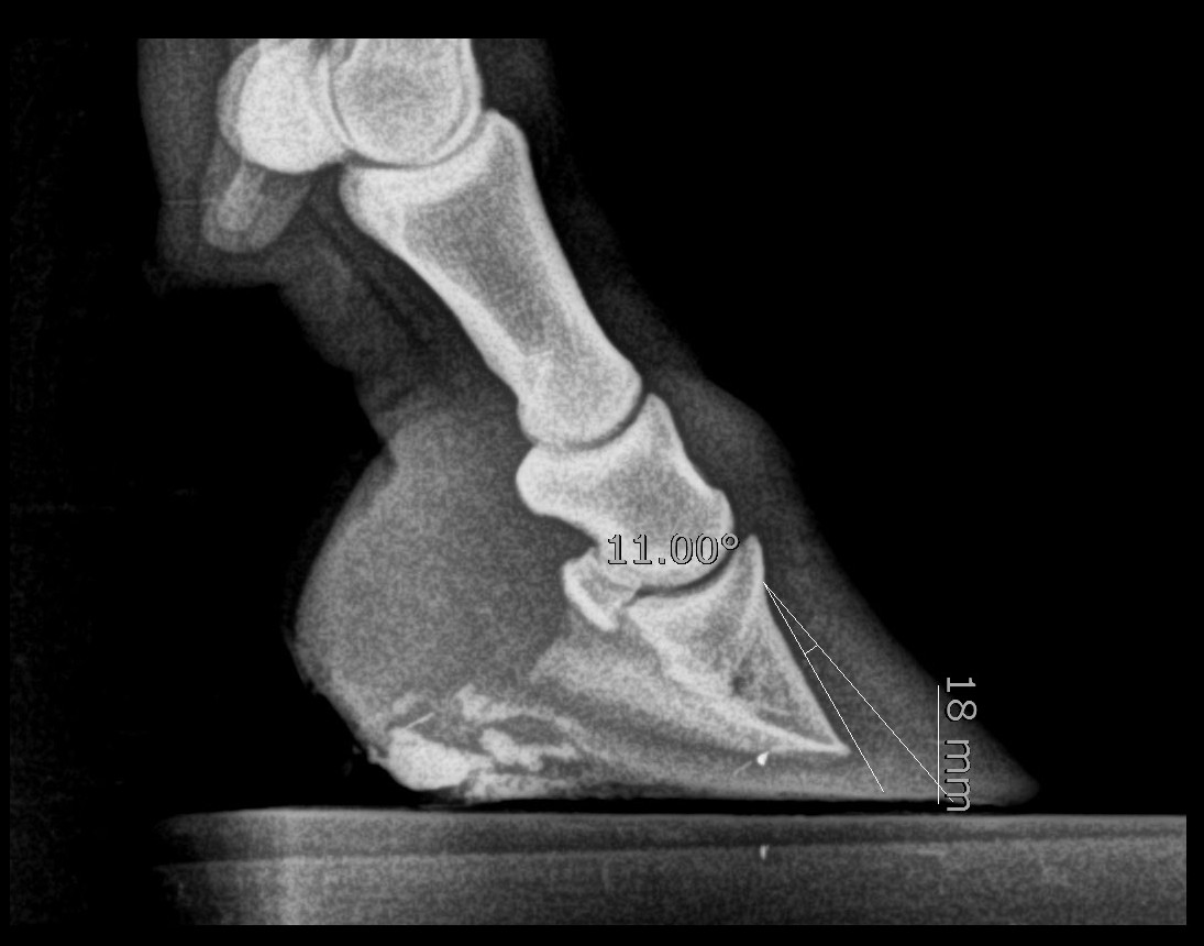
Rotation of the distal phalanx (P3) caused its tip to exert excessive pressure on the solar corium. This compression compromised the blood supply to the corium, limiting its capacity to produce new sole. As a result, sole depth was insufficient to adequately protect and support the distal phalanx, perpetuating a vicious circle in which the lack of sole allowed P3 to keep exerting excessive pressure on the solar corium and progressively worsening the biomechanical alterations of the hoof.
Initial assessment
A complete clinical examination was performed together with a radiographic study, in collaboration with his referring veterinarian, in order to determine the current state of the hoof and establish an individualised treatment plan.
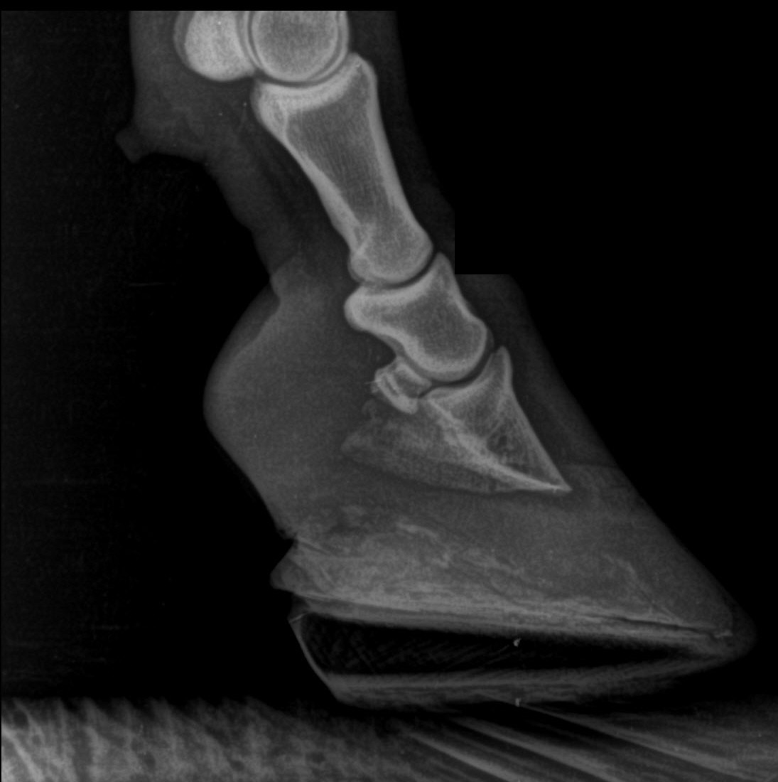
The radiographic assessment confirmed rotation of the distal phalanx (P3) and a markedly reduced sole depth, findings consistent with chronic laminitis.
Treatment
Custom 3D-printed horseshoes were designed and manufactured with the primary aim of reducing the tension exerted by the deep digital flexor tendon (DDFT) on the distal phalanx (P3). Decreasing this force reduced the pressure applied by the tip of P3 on the solar corium, restoring blood flow to this tissue and allowing sole production to resume.
The progressive increase in sole depth provided greater support to the distal phalanx, favouring its repositioning, creating a more favourable biomechanical environment for laminar repair and significantly improving the animal's comfort.
Results
From the first treatment cycle (6–8 weeks) an evident clinical improvement was observed, associated with the increase in sole depth and the reduction in pain.
After five consecutive treatment cycles, Mokka regained a physiological sole depth and the distal phalanx progressively returned to an appropriate anatomical position, findings confirmed by serial radiographic follow-up.
Laminar repair was evidenced both by the improved alignment of P3 and by the progressive normalisation of the white line during periodic trims, as well as radiographically. Clinically, the pony recovered normal locomotion, with resolution of the lameness and of the difficulty turning.
At the end of treatment, Mokka fully recovered his function and could be considered a case of clinical and radiographic resolution of laminitis, with absence of pain, adequate sole depth and a remarkable improvement in quality of life.